Inside the Ear: Exploring the Anatomy with an Animated 3D Model

Introduction Understanding the complex structure of the human ear can be challenging, especially when relying solely on textbooks or diagrams. However, with the advent of technology, we now have access to animated and 3D models that offer a more interactive and engaging way to explore the inner workings of the ear. In this blog, we’ll delve into the fascinating world of the ear canal using an animated 3D model, explaining its key components and how they work together to process sound and maintain balance.
Why Use an Animated 3D Model?
Traditional learning tools, like static images or physical models, often fall short in capturing the dynamic nature of how the ear functions. An animated 3D model provides a more comprehensive view by allowing users to:
- Visualize the Ear in Motion: See how sound waves travel through the ear canal, causing vibrations in the eardrum and how these vibrations are transmitted to the inner ear.
- Explore Each Part in Detail: Zoom in on specific components like the ossicles or cochlea, and view them from different angles to better understand their structure and function.
- Interact with the Model: Rotate, pan, and zoom into the model, offering a more hands-on experience that can enhance learning and retention.
Anatomy of the Ear: A Closer Look
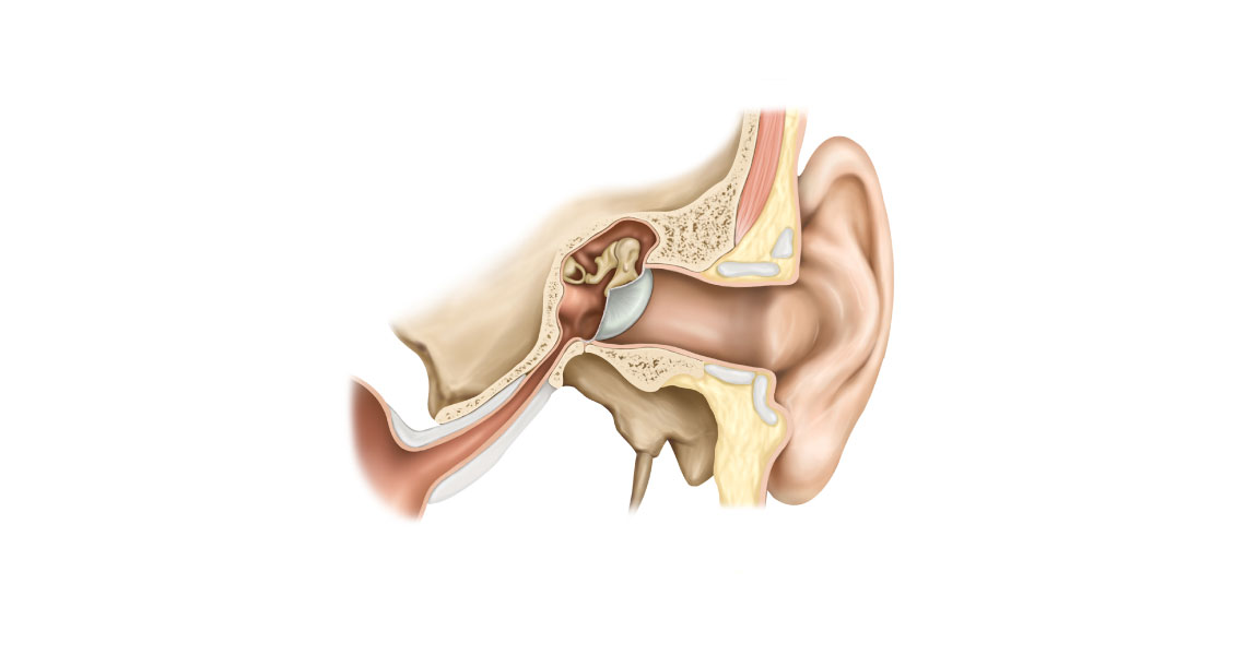
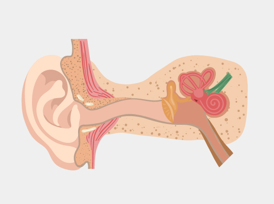
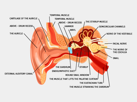
The Outer Ear
- Pinna (Auricle): The external, visible part of the ear that gathers sound waves and funnels them into the ear canal.
- Ear Canal (External Auditory Canal): The passage through which sound waves travel towards the eardrum.


The Middle Ear
- Eardrum (Tympanic Membrane): A thin membrane that vibrates in response to sound waves, initiating the process of hearing.
- Ossicles: The three smallest bones in the human body (malleus, incus, stapes) that amplify and transmit sound vibrations from the eardrum to the inner ear.
The Inner Ear
- Cochlea: : A spiral-shaped, fluid-filled organ that converts sound vibrations into electrical signals that the brain interprets as sound.
- Vestibular System: Comprising the semicircular canals and otolith organs, this system helps maintain balance by detecting head movements and changes in position.
- Auditory Nerve: The pathway that carries electrical signals from the cochlea to the brain, allowing us to perceive and recognize sounds.
The Benefits of Learning with 3D Models
Using a 3D model to explore the ear's anatomy offers several benefits:
- Enhanced Understanding: Seeing how each part of the ear interacts with the others in real-time provides a deeper understanding of its function.
- Improved Engagement: Interactive models are more engaging than traditional methods, making the learning process more enjoyable and memorable.
- Accessibility: : 3D models can be accessed from various devices, allowing students and educators to explore the ear’s anatomy anytime, anywhere.
Applications of 3D Ear Models in Education and Healthcare
Animated and 3D models of the ear are not just educational tools; they also have practical applications in healthcare:
- Medical Education: These models are invaluable for teaching medical students and professionals about ear anatomy and conditions affecting hearing and balance.
- Patient Education: Healthcare providers can use 3D models to explain ear-related diagnoses and treatment options to patients in a more understandable way.
- Surgical Planning: Surgeons can use detailed 3D models of a patient’s ear to plan complex procedures, improving precision and outcomes.
Conclusion
The human ear is a marvel of biological engineering, with each part playing a critical role in hearing and balance. Exploring the ear through an animated 3D model not only makes learning more interactive and accessible but also deepens our appreciation for the complexity of this vital organ. Whether you’re a student, educator, or healthcare professional, utilizing these models can greatly enhance your understanding of ear anatomy and function. Incorporating technology into learning and patient care represents the future of education and healthcare, offering new possibilities for how we explore and understand the world around us.